Rouviere’s Sulcus: A Guide to Safe Laparoscopic Cholecystectomy
Article information

Abstract
Purpose
Rouviere’s sulcus (RS) serves as an important anatomical landmark to avoid bile duct injuries during a laparoscopic cholecystectomy. However, there is significant paucity in literature regarding its surgical importance during laparoscopic surgeries. The aim of this study was to identify cases where RS was identified before dissection of the Calot’s triangle.
Methods
For this retrospective observational study, 500 patients who underwent a laparoscopic cholecystectomy at our hospital and who were operated on between September 2017 to August 2022 were reviewed. Identification of RS and its types were analyzed.
Results
Among all 500 cases, RS was present in 465 (93%) cases whereas it was absent in only 35 (7%) cases. RS was present in different forms of cholelithiasis. Open, closed, slit like, and scar type of RS was found in 75.29%, 12.90%, 3.21%, and 8.20%, respectively. Identification of RS along with achieving a critical view of safety in antero-superior direction to RS resulted in no injury to the bile duct in all 465 cases.
Conclusion
Identification of RS along with achieving a critical view of safety should be the aim in all laparoscopic cholecystectomy procedures. The RS is as an important landmark to reduce biliary tract injuries.
Introduction
The laparoscopic cholecystectomy is considered to be the gold standard operating procedure worldwide for the treatment of symptomatic gallstones. A laparoscopic cholecystectomy offers enhanced illumination and image quality provided by endoscopic cameras making visibility of this sulcus very clear. Although it is one of the most common surgical procedures, it has its own inherent limitations such as increased risk of injury to bile ducts [1]. Furthermore, it has been associated with more biliary, visceral, and vascular complications which are slightly higher when compared with open cholecystectomies [2,3].
To date, many surgical principles and approaches have been recommended to minimize the injuries which may occur to the bile duct. Patient safety is the utmost priority, therefore, it is important for the surgeons to identify the plane of the common bile duct before commencing dissection. A common landmark or reference point described in recent reports is the Rouviere’s sulcus (RS). The RS is a 2–5 cm fissure on the liver between the caudate process and right lobe.
The RS was not described during the traditional era of open cholecystectomy [4]. However, RS serves as an important anatomical landmark to avoid bile duct injuries during laparoscopic cholecystectomy [4–6]. There is a significant lack of studies in the literature regarding the surgical importance of locating RS during laparoscopic surgeries. The aim of this retrospective review was to identify cases where RS was identified before commencement of dissection of Calot’s triangle as a reference point to avoid thus preventing injury to the bile duct.
Materials and Methods
The current study was a retrospective, observational study conducted at JNUIMSRC, Jaipur, Rajasthan using medical records. The ethical clearance certificate (no.: JNUIMSRC/IEC/2023/20) was obtained from the University’s Ethics committee. It was in accordance with the Declaration of Helsinki of 1975.
A total of 500 patient cases with calculous cholecystitis between 20 years to 60 years of age who underwent a Laparoscopic Cholecystectomy (from September 2017 to August 2022) were reviewed. Known cases of laparoscopic cholecystectomy which converted to an open cholecystectomy were excluded from the study. In addition, patients with bleeding disorders, fatty liver, chronic liver disease, portal hypertension, cirrhosis, and infectious diseases of the liver; patients refusing consent and pregnant women were excluded from the study.
Before surgery patients had preoperative ultrasound scans, liver function tests, renal function tests, blood glucose level, coagulation profile, and complete blood count tests before the surgery. The operative procedure was laparoscopic cholecystectomy which was conducted under general anesthesia with the classical four port method. Cases where RS went undetected were noted for this retrospective review. The first port was a 10 mm infra-umbilical camera port, which was inserted directly after creating pneumo-peritoneum by CO2 insufflation via Veress needle. While the other three ports were inserted under direct vision. After visual exploration of the abdomen, the gallbladder was retracted up to the right axilla. Following which the Calot’s triangle was exposed with the lateral and inferior retraction of Hartman’s pouch. The frequency and the type of the RS were tabulated. Broadly, the types of RS are described as the open type where a right hepatic pedicle is seen and the sulcus is open throughout its length. The closed (fused) type is where the pedicle cannot be visualized or the sulcus only opens at its lateral end, and the absent type is where the sulcus cannot be identified during the operation and the slit type is when the sulcus is narrow and shallow. The RS is rarely closed (fused) and is represented by a white line of fusion (scar like).
Once the RS was identified, dissection commenced. The dissection of cystic duct and cystic artery in the Calot’s triangle was based on the principles of the critical view of safety in antero-superior direction to RS. The cystic duct and cystic artery were then clipped and cut. Dissection of the gallbladder from its bed was performed using electrocautery and blunt dissection, until completely removed under vision from epigastric port. After the insertion of a tube drain in the sub hepatic region, the sites of the ports were closed. Any significant intraoperative or postoperative event was also recorded.
Results
All patients presented to the Outpatient Department of Surgery with symptomatic cholelithiasis. Patients were predominantly females (71.2%) and the average age of the study sample was 36.23 ± 9.05 years with a median of 33. Female to male ratio in the current study sample was 2.49:1.
A preanesthetic check-up for fitness for surgery was conducted for all patients. The average duration of surgery was 46.30 minutes (20–75 minutes) and the average hospital stay was 2.43 days (2–5 days). Among the 500 operated cases, RS was found to be present in 465 (93%) cases. Among the 465 cases where RS was identified, no injury to the bile duct was reported after the critical view of safety (CVS) was achieved. Thus, the RS was reported to be present in overwhelming majority (Table 1).
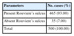
Incidence of Rouviere’s sulcus.
Among these 465 cases, morphologically different varieties of RS were observed such as open, closed, slit or scar like sulcus. Furthermore, deep sulcus was categorized as open type in 75.3% (350 cases), and closed (fused) type in 12.9% (60 cases; Table 2).

Comparative incidence of the types of Rouviere’s sulcus.
Discussion
Laparoscopic removal of the gallbladder or cholecystectomy has become the most common procedure of choice for routine removal of gallbladder. Based on an increased need and frequency of laparoscopic cholecystectomies, the incidences of bile duct injuries have also increased. To minimize these injuries, the identification of common bile duct along with a critical view of safety should be encouraged. Many suggestions and recommendations have been proposed so far and one of these is an extrabiliary landmark RS, which makes the identification of the target tissue easier for dissection. The most significant factor that results in bile duct injury during a laparoscopic cholecystectomy was reported to be spatial disorientation on the part of the surgeon [2]. To avoid this, they used the RS as an extrabiliary fixed point to start dissection and was found to be a useful anatomical landmark for this purpose [2]. Surgeons have come to rely on these landmarks (RS and CVS) for preventing bile duct injuries. Thus, the present study was conducted to describe the surgical importance of RS during laparoscopic surgeries.
In the current study sample, patients were predominantly females, a total of 356 out of 500 cases. These results were in accordance with the studies conducted by Jha et al [7], and Rosen and Broody [8]. RS was present in different forms in 465 cases (93%) of cholelithiasis in our study. The open type of sulcus was found in 75.29% of patients which was similar to Jha et al [7] (63.33%), Singh et al [9] (71%), and Zubir et al [10] (68.13%) [11]. However, the sulcus was not seen/absent in 35 cases (7%). In cases where the RS is not seen, it is recommended that the surgeon should rely on other anatomical landmarks such as the umbilical fissure and segment 4 of the liver.
Identification of the RS in the beginning and keeping a track of it during the operation can be learned by trainee surgeons. This helps them to avoid the wrong plane and gives a fixed point from the beginning. In addition, a panoramic view of the surgical field provides a better view of these clues/landmarks to the surgeon and helps them perform a safe cholecystectomy.
Conclusion
Identification of RS along with achieving a CVS should be the aim in all laparoscopic cholecystectomy procedures. The results of this retrospective review of the patient charts shows the importance of RS as a landmark to reduce biliary tract injuries.
Notes
Author Contributions
Conceptualization: RP and LM. Methodology: RP and LM. Formal investigation: RP, LM, Ghaus MC and AT. Data analysis: LM, GMC and AT. Writing original draft: RP, LM and GMC. Writing - review and editing: RP, GMC and AT.
Conflicts of Interest
The authors have no conflicts of interest to declare.
Ethical Statement
This research did not involve any human or animal experiments.
Data Availability
All relevant data are included in this manuscript.
Funding
None.