

• KSACS
  • KTACN
  • Contact us
  • E-Submission
ABOUT
BROWSE ARTICLES
JOURNAL POLICIES
FOR CONTRIBUTORS

Articles

Interest Image

Hemodynamically Stable Patient after Inferior Vena Cava Penetrating Injury by Stab Wound

찬규 이, M.D., 재훈 김, M.D., 길환 김, M.D.
Journal of Acute Care Surgery 2017;7(2):92-93.
Published online: October 30, 2017

Jae Hun Kim, M.D. Department of Trauma Surgery, Pusan National University Hospital, 179 Gudeok-ro, Seo-gu, Busan 49241, Korea Tel: +82-51-240-7369, Fax: +82-51-240-7719, E-mail: jjangmdkmdk@hanmail.net
• Received: September 13, 2017   • Revised: September 28, 2017   • Accepted: September 28, 2017

Copyright: © 2017 by Korean Society of Acute Care Surgery

This is an Open Access article distributed under the terms of the Creative Commons Attribution Non-Commercial License (http://creativecommons.org/licenses/by-nc/4.0) which permits unrestricted non-commercial use, distribution, and reproduction in any medium, provided the original work is properly cited.

  • 5,578 Views
  • 35 Download
prev
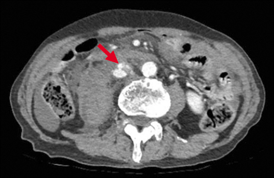
74세 남자 환자가 자살 목적으로 회칼로 자신의 복부를 수상하여 응급실을 내원하였다. 내원 시 활력징후는 안정적이었으며 이학적 검사상 배꼽 주변의 4 cm 정도의 개방성 자상을 보였다. 복부 전산화단층촬영상 장간막에 출혈 및 하대정맥에 혈관 외 유출 소견을 보여 응급수술을 시행하였다. 수술 소견에서 장간막에 출혈과 소장의 손상이 보여 혈관 결찰술과 소장 절제술을 시행하였다. 복부 전산화단층촬영에서 하대정맥은 신장정맥의 분지 부위 하방에 혈관 전벽 손상이 의심되었으나(Fig. 1), 후복강에서 약 2 L 가량의 출혈과 하대정맥의 전벽을 역 Y자 모양으로 관통하여 후벽까지 포함하는 손상이 발견되었다. 스폰지스틱(sponge stick)으로 손상 부위 근위부 및 원위부를 눌러 혈류를 차단한 후, 후벽은 전벽 손상을 벌려서 단순연속봉합으로, 전벽은 이중으로 단순연속봉합을 사용하였다(Fig. 2, 3). 환자는 아스피린(aspirin)을 단독 복용하며 외래 추적 관찰을 하였고 수술 3개월 후 전산화단층촬영 검사상 하대정맥의 협착 및 혈전 없이 회복을 보였다(Fig. 4). 복부 외상 환자 중 진단적 개복술을 받은 환자의 4% 미만에서 하대정맥손상이 발견되며, 이들은 60% 가량의 사망률을 보인다[1,2]. 하대정맥 손상은 빠른 수술적 치료가 중요하며 대부분 결찰 또는 일차봉합술이 수술방법이다.
Fig. 1
Abdominal computed tomography showed extravasation at inferior vena cava anterior wall (arrow).
ACS_07_092_fig_1.jpg
Fig. 2
Abdominal computed tomography showed extravasation at inferior vena cava anterior wall (arrow).
ACS_07_092_fig_2.jpg
Fig. 3
Pattern diagram of anterior and posterior inferior vena cava wall.
ACS_07_092_fig_3.jpg
Fig. 4
Abdominal computed tomography in three month after surgery showed that the inferior vena cava were not narrowed (arrow).
ACS_07_092_fig_4.jpg
본 연구는 2016년도 부산대학교병원 임상연구비 지원으로 이루어졌음.

Conflicts of Interest

No potential conflict of interest relevant to this article was reported.

  • 1. Maciel JD, Plurad D, Gifford E, deVirgilio C, Koopmann M, Neville A, et al. Predictors of mortality in patients with penetrating inferior vena cava injuries surviving to the operating room. Am Surg 2015;81:1000–4.
  • 2. Fonseca AZ, Ribeiro MA Jr. Penetrating injury to the inferior vena cava. Surgery 2015;157:1174–5.

Figure & Data

References

    Citations

    Citations to this article as recorded by  

      Download Citation

      Download a citation file in RIS format that can be imported by all major citation management software, including EndNote, ProCite, RefWorks, and Reference Manager.

      Format:

      Include:

      Hemodynamically Stable Patient after Inferior Vena Cava Penetrating Injury by Stab Wound
      J Acute Care Surg. 2017;7(2):92-93.   Published online October 30, 2017
      Download Citation
      Download a citation file in RIS format that can be imported by all major citation management software, including EndNote, ProCite, RefWorks, and Reference Manager.

      Format:
      • RIS — For EndNote, ProCite, RefWorks, and most other reference management software
      • BibTeX — For JabRef, BibDesk, and other BibTeX-specific software
      Include:
      • Citation for the content below
      Hemodynamically Stable Patient after Inferior Vena Cava Penetrating Injury by Stab Wound
      J Acute Care Surg. 2017;7(2):92-93.   Published online October 30, 2017
      Close

      Figure

      • 0
      • 1
      • 2
      • 3
      Hemodynamically Stable Patient after Inferior Vena Cava Penetrating Injury by Stab Wound
      Image Image Image Image
      Fig. 1 Abdominal computed tomography showed extravasation at inferior vena cava anterior wall (arrow).
      Fig. 2 Abdominal computed tomography showed extravasation at inferior vena cava anterior wall (arrow).
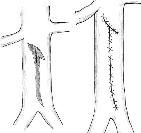
      Fig. 3 Pattern diagram of anterior and posterior inferior vena cava wall.
      Fig. 4 Abdominal computed tomography in three month after surgery showed that the inferior vena cava were not narrowed (arrow).
      Hemodynamically Stable Patient after Inferior Vena Cava Penetrating Injury by Stab Wound
      TOP