• KSACS
  • KTACN
  • Contact us
  • E-Submission
ABOUT
BROWSE ARTICLES
JOURNAL POLICIES
FOR CONTRIBUTORS

Articles

Interesting Image

Retrosternal Hemorrhage after Cardiac Compression

Journal of Acute Care Surgery 2018;8(1):40-41.
Published online: April 30, 2018

Division of Trauma Surgery, Department of Surgery, Ajou University School of Medicine, Suwon, Korea

Yo Huh, M.D. Division of Trauma Surgery, Department of Surgery, Ajou University School of Medicine, 164 WorldCup-ro, Yeongtong-gu, Suwon 16499, Korea Tel: +82-31-219-7764, Fax: +82-31-219-7781, E-mail: ermdhuhyo@gmail.com ORCID: http://orcid.org/0000-0002-1220-1534
• Received: December 14, 2016   • Revised: March 13, 2017   • Accepted: March 15, 2017

Copyright: © 2018 by Korean Society of Acute Care Surgery

This is an Open Access article distributed under the terms of the Creative Commons Attribution Non-Commercial License (http://creativecommons.org/licenses/by-nc/4.0) which permits unrestricted non-commercial use, distribution, and reproduction in any medium, provided the original work is properly cited.

  • 6,197 Views
  • 39 Download
prev
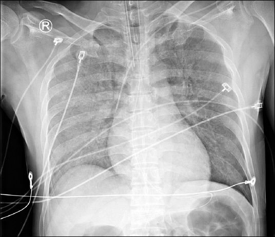
A 43-year-old male visited the Trauma Center after a pedestrian traffic accident. During initial resuscitation, suddenly the ventricular fibrillation was detected. Cardiac compression and cardioversion were performed for about 10 minutes, and his rhythm was recovered. A chest radiograph was taken (Fig. 1), and focused assessment with sonography in trauma (FAST) was negative (Pericardial effusion wasn’t seen, and heart movement was decreased, However it was difficult to distinguish from the effect of ventricular fibrillation). He was admitted to the intensive care unit and a transesophageal echocardiography (TEE) was performed for cardiac evaluation (Unfortunately, the image wasn’t saved). TEE presented cardiac tamponade features, hence a large amount of fluid was administrated instead of using more inotropic drug. Computed tomography (CT) scan was taken and the CT showed retrosternal hematoma which compressed the right ventricle (Fig. 2). Because retrosternal hematoma was not easily detected in FAST, CT or TEE could be considered for evaluation after cardiac compression [1,2].
Fig. 1
Chest radiograph showed normal cardiac contour.
ACS_8_040_fig_1.jpg
Fig. 2
(A) Indicator showed active bleeding of retrosternal area. (B) Heart was compressed by retrosternal hematoma, and both lung atelectasis was shown
ACS_8_040_fig_2.jpg
No potential conflict of interest relevant to this article was reported.
  • 1. Hsu LW, Chong CF, Wang TL, Wu BH. Traumatic mediastinal hematoma: a potentially fatal condition that may be overlooked by traditional focused assessment with sonography for trauma. Am J Emerg Med 2013;31:262e1–3.
  • 2. Kim YH, Kwon JB, Park CB, Choi SY. Extrapericardial cardiac tamponade by a retrosternal haematoma after blunt chest trauma. Eur J Cardiothorac Surg 2012;41:958.

Figure & Data

References

    Citations

    Citations to this article as recorded by  

      Download Citation

      Download a citation file in RIS format that can be imported by all major citation management software, including EndNote, ProCite, RefWorks, and Reference Manager.

      Format:

      Include:

      Retrosternal Hemorrhage after Cardiac Compression
      J Acute Care Surg. 2018;8(1):40-41.   Published online April 30, 2018
      Download Citation
      Download a citation file in RIS format that can be imported by all major citation management software, including EndNote, ProCite, RefWorks, and Reference Manager.

      Format:
      • RIS — For EndNote, ProCite, RefWorks, and most other reference management software
      • BibTeX — For JabRef, BibDesk, and other BibTeX-specific software
      Include:
      • Citation for the content below
      Retrosternal Hemorrhage after Cardiac Compression
      J Acute Care Surg. 2018;8(1):40-41.   Published online April 30, 2018
      Close

      Figure

      • 0
      • 1
      Retrosternal Hemorrhage after Cardiac Compression
      Image Image
      Fig. 1 Chest radiograph showed normal cardiac contour.
      Fig. 2 (A) Indicator showed active bleeding of retrosternal area. (B) Heart was compressed by retrosternal hematoma, and both lung atelectasis was shown
      Retrosternal Hemorrhage after Cardiac Compression
      TOP