심폐소생술로 인해 발생한 대동맥박리
Aortic Dissection Following Cardiopulmonary Resuscitation
Article information
J Acute Care Surg. 2016;6(1):40-41
Publication date (electronic) : 2016 April 30
doi :
https://doi.org/10.17479/jacs.2016.6.1.40
Received 2016 April 07; Revised 2016 April 12; Accepted 2016 April 14.
Body
기저질환이 없는 54세 남자 환자가 위암으로 진단 후 위절제술 시행예정이었다. 수술실에 입실하여 마취 전 예방적 항생제 cefotetan을 정맥 주입한 후 아나필락시스로 추정되는 심정지가 발생하여 26분간 수술실에서 심폐소생술이 지속되었다. 심폐소생술 중 시행한 경식도 심초음파에서 좌심실의 무수축 상태가 지속되어 즉시 체외막산소화장치를 적용하고 자발순환회복이 확인되어 56분만에 중환자실로 이동하였다. 5시간 후 재시행한 경흉부 심초음파에서 59%의 심박출계수로 좌심실 수축 소견이 정상적으로 회복되고 혈류역학적으로 안정화되어 9시간만에 체외막산소화장치를 제거하였다. 아나필락시스 쇼크에 의한 심폐소생술 후 발생한 급성폐손상과 급성신손상 등으로 집중치료 25일째 일반병실로 전동되었으며 다행히 심폐소생술에 의한 신경학적 손상 소견은 없었다. 심정지 발생 20일째, 향후 위암에 대한 치료결정을 위해 시행한 컴퓨터 단층촬영 검사에서 좌측 4번째, 5번째 늑골 골절과 함께 대동맥박리가 진단되었다(Fig. 1, 2). 대동맥박리는 하향 흉부대동맥에서부터 신동맥 상방까지 침범하는 DeBakey III형으로 혈압조절을 포함한 약물치료를 시행하면서 추적관찰 중이다[1,2].

Transverse section of computed tomography.
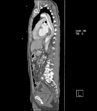
Sagittal plane of aortic dissection computed tomography.
References
1. Oren-Grinberg A, Shahul S, Sarge T. Dissection of the thoracic aorta following cardiopulmonary resuscitation. Crit Ultrasound J 2011;3:25–7.
10.1007/s13089-011-0056-5.
2. Okuda T, Takanari H, Shiotani S, Hayakawa H, Ohno Y, Fowler DR. Pericardial tear as a consequence of cardiopulmonary resuscitation (CPR) involving chest compression: a report of two postmortem cases of acute type A aortic dissection with hemopericardium. Leg Med (Tokyo) 2015;17:201–4.
10.1016/j.legalmed.2014.12.001. 25533925.
Article information Continued
Copyright: © 2016 by Korean Society of Acute Care Surgery